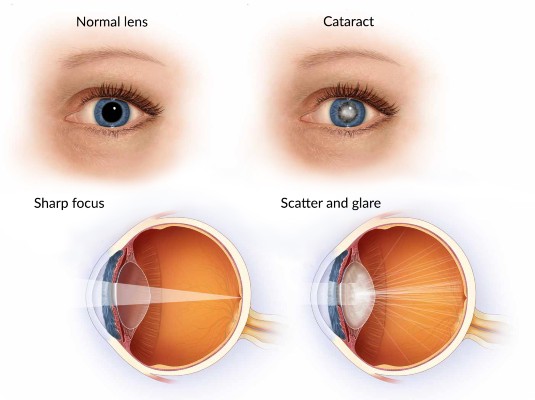
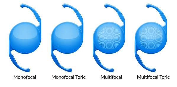
What is it?

Cataract refers to clouding changes in the clear lens in the eye. The lens is situated just behind the iris, which is the coloured part of the eye. More…

Image: Cataract

See More Information

In a healthy eye, light passes though the cornea, the clear part at the front of the eye, before it passes through the pupil, which is the black dot in the centre of the iris. The pupil is a hole in the iris, and it changes size to control how much light enters the eye.
The lens is located immediately behind the pupil and focuses light onto the light sensitive layer at the back of the eye.

Age related changes to the transparency of the lens are responsible for the majority of cataracts. These changes usually progress slowly and result in uniform clouding of the lens over a long time. However, cataracts do not all appear the same or progress at the same rate. In some cases, they may progress rapidly, leading to visual impairment in a matter of months.

Nuclear cataract- This type of cataract is the result of ageing and is the most prevalent, responsible for over half of all cataracts.

Cortical cataract – White opacities form in the outer part of the lens and gradually progress to the centre where they produce symptoms.

Subcapsular cataract – This cataract begins at the back of the lens where it appears like frosting that gradually spreads out. Initially it produces glare, particularly noticeable when driving at night. Eventually it causes significant vision loss, often in a relatively short space of time Cataracts will progress and, if neglected, will eventually reduce the vision to the point where it is no longer possible to read or safe to drive.
There is no preventative treatment for cataracts although some lifestyle modifications may delay the onset of symptoms. These include general measures that have been encouraged to reduce the risk of heart disease and skin cancer, namely, reduction of alcohol intake, stop smoking, healthy diet that includes fruits and vegetables, good diabetic control and the use of sun protection (sunglasses).

How do you get it?

Cataracts are usually caused by ageing changes in the lens. Other causes are listed here… The proteins in the lens are precisely arranged into a structure that makes it transparent. In time the proteins start to break apart and the orderly arrangement becomes disrupted, which leads to loss of transparency.

These changes start to become apparent after the age of 60 and steadily progress as we get older. By the age of 70 about 20% of people will have cataracts. This increases to about 50% by the age of 80. There is a slightly higher rate of cataracts in women.

In addition to ageing the following factors may also contribute to cataract formation
  • Family members with cataracts
  • Certain medical conditions, such as Diabetes
  • Trauma to the eye
  • Eye surgery
  • Smoking
  • Sun exposure without sunglasses
  • Radiation to the head or upper body
    • Certain medications, such as corticosteroids
Do I have it?

Cataracts grow slowly, over months, or more commonly, years. Initially there may be some changes in the lens, but they cause no symptoms at this stage. There is no vision loss, and the eye appears completely normal. Only slit lamp examination will reveal these early changes.

Gradually the vision will become hazier and will not improve with glasses. Colours may change, with more yellows and fewer blues, but this may not be obvious as the changes occur slowly.

Bright lights may cause glare and all colours appear slightly washed out. Nuclear cataracts may also increase short sightedness, improving near vision. Unfortunately, this change is temporary and indicates that the cataract is becoming more significant, and soon the vision at all distances will deteriorate.

As soon as you notice any reduction in your vision, you should see your local optometrist.

They will check your prescription and test your vision. They will also perform an eye examination to exclude other eye problems. If cataracts are found, you will be referred to an eye surgeon (ophthalmologist) who will confirm the diagnosis and explain how your vision can be restored.

How is it treated?

Cataracts are irreversible and treatment is surgical.

A new pair of glasses will not compensate for the loss of transparency in the lens. There is no diet or drop or exercise that restores clarity to the lens. More information about cataract surgery…

The cataract operation

Cataract surgery is performed under local anaesthetic with sedation.
In practice this means that the anaesthetist will provide you with intravenous sedation that
puts you to sleep for a few minutes while a local anaesthetic is applied to your eye socket.
When you wake up you will be relaxed and comfortable with no sensation around the eye, and you will be taken into theatre.

A small device, called a speculum, is placed to keep the lids open. Your eyelids and the contents of your eye socket will be numb, and you will feel no discomfort. You will be able to keep blinking normally with the other eye, or keep it closed.

The operation starts with a small incision through the edge of the cornea to provide access to the space between the clear cornea and the coloured iris. This space is called the anterior chamber. Once in the anterior chamber the operation is performed through the dilated pupil.

The cataract is located directly behind the iris and is held in place by a very thin membrane called a capsule that wraps around the lens. This capsule is important, as it will also provide support for the intraocular lens after the cataract has been removed.
The capsule, in turn, is attached to the wall of the eye (ciliary body) by means of fine fibrous strands (zonules), like springs on a circular trampoline.

An opening is made through the front of the capsule to allow access to the cataract. A tiny metallic probe, which vibrates at ultrasonic frequency is then used to break the cataract into very small pieces. These pieces are vacuumed out of the eye by means of a specialised instrument called a phacoemulsification handpiece.

Once the cataract has been removed the capsule is cleaned and is now ready to receive the intraocular lens (IOL). The hole in the capsule is too small for the IOL to fit though directly, and it needs to be rolled up, like a scroll, and posted through the opening.

Once inside the capsular bag, it slowly unfolds and resumes the role of focusing light onto the back of the eye.
The corneal incisions are less than 3mm wide and are designed to be self-sealing. No stitches are required.

A small amount of antibiotic is placed in the anterior chamber at the end of the procedure and a transparent shield is placed over the eye for mechanical protection.

The operation takes about 10 minutes.

Aftercare

The shield will stay on the eye overnight.
You will be seen at the practice the following day where the nurse will remove the shield and check your vision. She will also explain everything that you need to know regarding aftercare.

You will be seen by your ophthalmologist to confirm that the eye and vision are stable.

The shield will need to be reapplied at night for a week after the surgery.

You will have a prescription for drops to apply to the operated eye. These drops are used four times daily for four weeks.
Most patients will be booked for surgery to both eyes. However, the procedures will not be performed on the same day. Surgery for the second eye is usually planned for 2 weeks after the first.

If you would like to see an ophthalmologist to arrange an assessment contact our staff on (07) 5506 6777.

The cataract has to be removed and replaced by an implant that performs the same function. This is known as an intraocular lens (IOL) implant. It is made from a variety of flexible, transparent materials that do not degrade and it will never wear out. More information on intraocular lenses…

Intraocular lenses
The intraocular lens is made from a type of plastic that is moulded or cut provide a lens of a particular strength. Every eye is unique and the strength of the IOL that is required will vary from patient to patient. A range of strengths is available to allow the most suitable lens to be selected for each eye. There are numerous manufacturers that provide IOLs and most will also have a selection of lens types. The type of lens that is best suited to your eye will depend on your biometry (a list of eye measurements made prior to surgery) as well as personal preference and I we will explore these lens types below. An IOL is selected primarily to provide clear distance vision. All the IOLs will achieve this. An IOL that provides only distance vision is referred to as monofocal, meaning that it only focuses on one place (distance), which for practical purposes means everything further than about 2 or 3 meters away. Anything closer that this will become increasingly blurred. In addition, some IOLs will provide intermediate or near vision as well, and these may be referred to as multifocal IOLs. There is also an option to correct astigmatism with an intraocular lens known as a toric lens. A normal cornea has a uniform curvature, like a soccer ball. A cornea with astigmatism is more curved in one direction than the other, like a rugby ball. A cornea with astigmatism is unable to focus light sharply unless the light passes through a second lens that can correct the astigmatism. This is effectively what the optometrist does when prescribing glasses. The correcting lens can also be the toric intraocular lens that is placed at the time of surgery. The advantage of this approach is that good vision is now possible without glasses. Broadly speaking the monofocal IOL will have the lowest risk of visual disturbance after cataract surgery. Multifocal IOLs have concentric grooves on the surface which allows the lens to focus over a larger range. However, this design introduces the possibility of visual disturbance in the form of glare and halos. The halos are visible when looking at lights at night and vary from barely noticeable to significant. It appears that a greater the degree of near vision correction correlates to a greater risk of visual disturbance.

Image: IOL Range

Cataract surgery is one of the most frequently performed operations worldwide and 99.5% of patients have no complications. However, there is always a small risk with any procedure, and this will be discussed with you in detail before you decide to proceed with surgery.