A range of instruments are used to measure and record the function and condition of the eye, and this information is used to diagnose ocular problems as well as monitor the effectiveness of their treatment.
FUNDUS CAMERA
The fundus camera takes a photograph of the structures at the back of the eye (called the fundus). The photograph is the same image that your ophthalmologist sees when they look through the pupil, into the eye. The optic nerve, blood vessels and retina may all be affected by ocular disease and a fundus photograph is a valuable tool to help with diagnosis and monitoring of these conditions.
Fundus Camera
Fundus Photography
AUTOREFRACTION

Autorefraction will measure your eyes to provide a fast and reasonably accurate spectacle prescription.

If you are far sighted, near sighted or have astigmatism, you may have trouble seeing well without a pair of glasses.
When you go to see your optometrist, they test your vision by putting a range of lenses in front of your eyes until they can compensate for your condition. They fine tune this until they have the perfect combination of lenses required to provide the best vision. This is referred to as your prescription.
An autorefractor is not as accurate as an optometrist but is able to provide a reasonably accurate measurement of your prescription. This is particularly useful for patients who do not wear glasses or those who have not seen an optometrist for a long time.

AUTOREFRACTION
LENSMETER
A lensmeter can measure a pair of glasses in order to determine their prescription. Patients who wear glasses generally don’t know what their prescription is. There is really little reason to know, as long as the glasses do what they are supposed to. However, your ophthalmologist needs to know. When you are referred by an optometrist, they will usually include your prescription in the correspondence. If the referral does not contain your prescription, we are able to measure your glasses with a lensmeter in order to obtain it.
LENSMETER
VISUAL FIELD ANALYSER

A visual field analyser maps out your visual field. It monitors areas in your visual field that function poorly.

When you look at something, you are usually focussed on an object in the centre of your vision. This object is in sharp focus and this is what you are concentrating on.
Let us use an example that involves driving a car. You may be focussed on the car in front of you, but you are aware of a lot more than the car. You can see all the other cars in front of you, the road below, the sky above and everything in between, even though you are not focused on them specifically. Everything that you perceive visually is called your visual field.
When we test your vision on the chart, we are testing your central vision. Sometimes we need to also test your peripheral vision and we do this with a visual field analyser. It is not a passive test, like many of the other investigations. You need to respond by pushing a button when you see a light flash. It may take a while to test your entire visual field, but the results are valuable in diagnosis and monitoring of a range of ocular and neurological conditions.

VISUAL FIELD ANALYSER
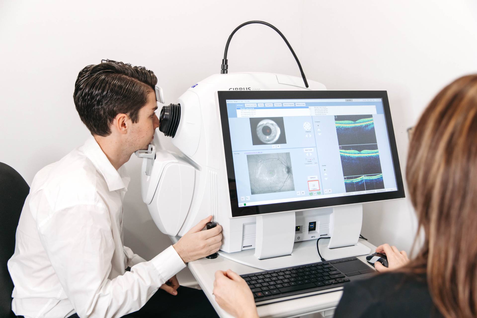
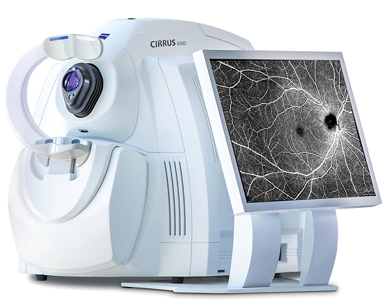
OCULAR COHERENCE TOMOGRAPHY (OCT)

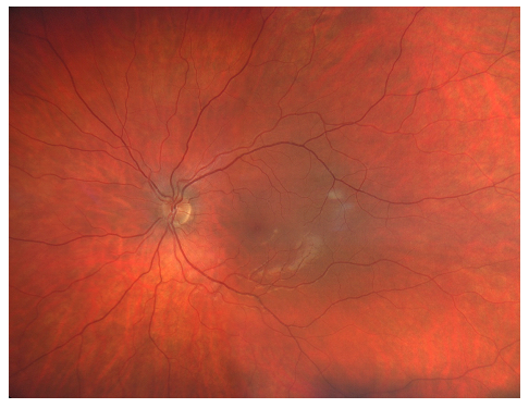
The OCT provides a highly detailed view of the internal structure of the retina.

The structures in the eye that lie along the path of light, from the cornea at the very front of the eye, to the retina, at the back, all share a very unique feature for living tissue. They are transparent.
We have been able to develop technologies that use light to look at the retina (which is also transparent) in incredibly fine detail. Almost at cellular level. This has been a massive boost to our study of eye disease and, even more significantly, to our management of eye disease.
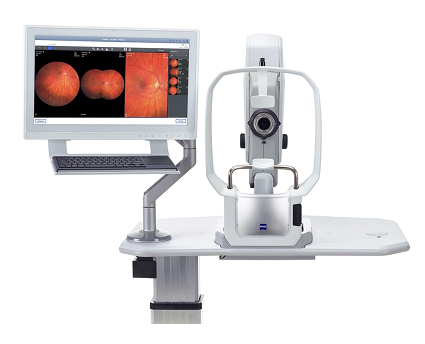
The new models of OCT, like the one in our practice, which is illustrated below, analyses the flow of blood through vessels in the retina. There are several conditions, including diabetic eye disease, where this information is important in determining treatment and providing a prognosis.

OCT (Optical Coherence Tomography)
TONOMETRY

A tonometer is a device that measures the pressure in the eye.

Fluid is constantly produced in small quantities inside the eye. This fluid then circulates until it reaches a structure through which it can drain out of the eye and into the bloodstream. This additional fluid results in a slight pressure in the eye (like blowing a bit of air into a balloon). The speed at which fluid is able to drain from the eye has an effect on the pressure. The pressure will go up if the fluid encounters resistance to draining.
If the pressure gets too high it results in damage to the optic nerve, which sends the images from the retina to the brain. High pressure in the eye is not painful. In fact, you won’t know that your pressure is high until your optometrist or ophthalmologist tell you it is.
The instrument they use to check this pressure is called a tonometer.

TONOMETRY
BIOMETRY

This instrument is used for patients that are planning to have cataract surgery.

A cataract is simply the lens in the eye that is no longer transparent. The cataract needs to be removed and replaced by a clear, artificial lens that can perform the same function.
This is called an intraocular lens (IOL) and it is manufactured in a range of strengths. We need to determine which IOL strength you need, and we do this by recording specific measurements in your eye. These measurements are referred to as biometry and this is the instrument that is used to record the data and calculate the required IOL strength that will provide you with clear distance vision after cataract surgery.

BIOMETRY